
"Keppler's Vault 72: The Eye" revisits a March 1961 Modern Photography article, "The Eye: From Lens to Darkroom" by George Wald, which explores how the human eye compares to a photographic lens. The blog author, who has lived with poor vision his whole life, frames the piece around his personal curiosity about how the eye achieves such depth of field and low-light sensitivity compared to a camera lens, wondering what f/stop or ASA equivalent the human eye might have.
The original article details the parallels and differences between eyes and lenses: both use an iris to regulate light and a lens to focus and invert images, but while film captures light on a flat plane, the eye relies on a curved surface of rods and cones sending signals to the brain. The piece explains how cones handle bright, colorful vision while rods take over in dim light at the cost of color detail, likening this shift to switching from vibrant color film to fast black-and-white film. It also touches on how other animals' eyes work differently, citing pinhole-like vision, compound eyes such as houseflies', and the scanning eye of the Copilia. The article further contrasts how focus and aberration correction are achieved differently in eyes (via shape change and variable refraction) versus lenses (via moving elements and coatings), and describes an experiment involving red and blue lights to demonstrate shifting color sensitivity between rods and cones. It even recounts a 19th-century experiment by physiologist Willy Kuehne involving decapitated rabbits to show retinal image formation.
The blog author found the chemical discussions less engaging and notes that the article never actually answers his original question about the eye's equivalent f/stop or film speed, but still calls it a fascinating read. He closes by speculating about how far eye-related optical research has advanced since 1961 and whether it could inspire future camera technology—such as lenses that change shape for focus, use variably refractive materials, or sensors capable of variable ISO within a single image—noting that many modern lenses still rely on century-old designs like the Tessar and Heliar formulas.
AI-generated summary. May not capture every detail — read the full article below for the complete picture.
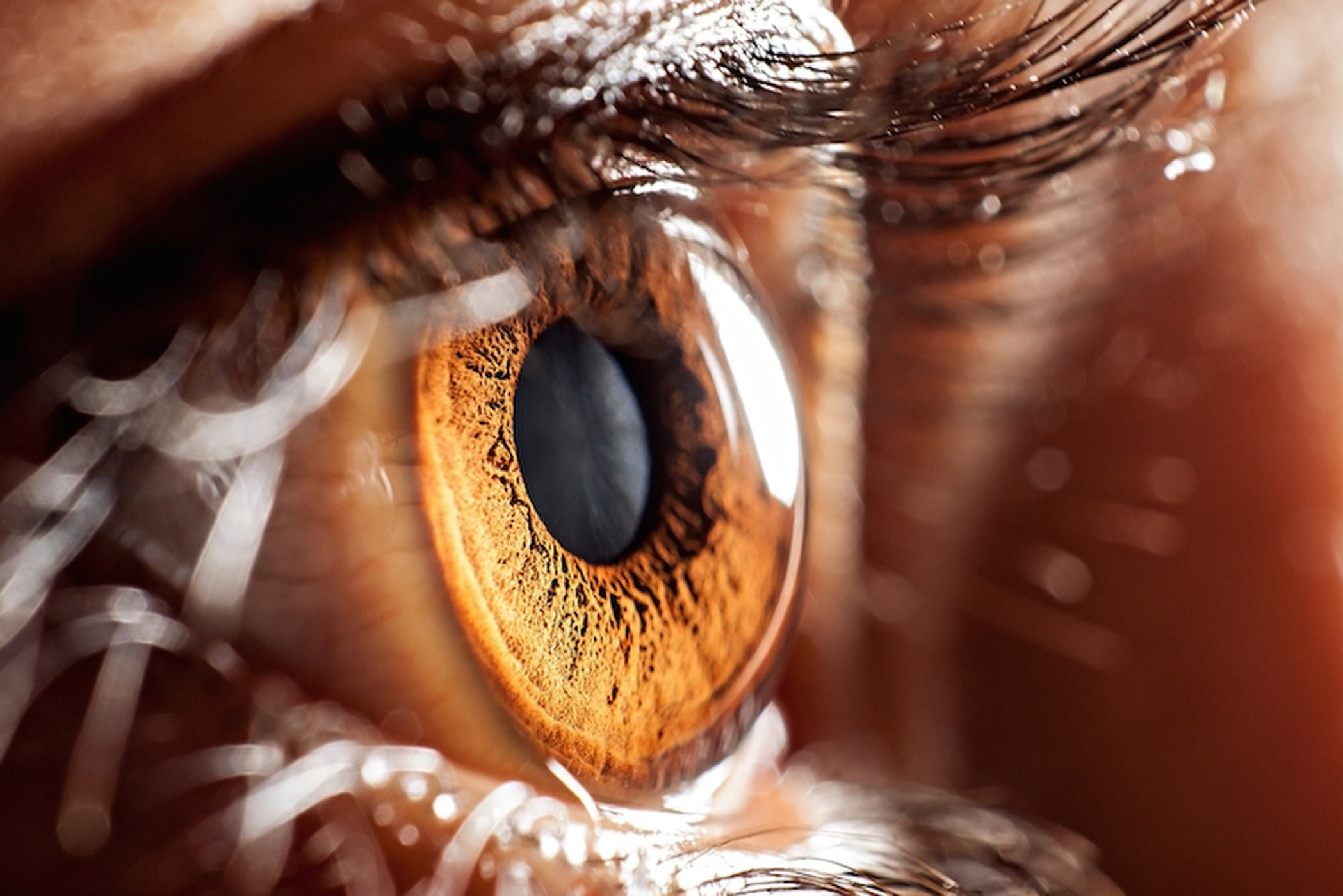 For this site’s first post on the human anatomy, I bring you an interesting article from the March 1961 issue of Modern Photography called “The Eye: From Lens to Darkroom” in which author George Wald investigates how our own eye compares to that of an optical lens.
I’ve suffered from poor vision my entire life, being born without sight in my right eye. I also have severe nearsightedness, astigmatism, and nystagmus in my left so I’ve always had a bit of a fascination with how the eye works.
As a photographer with only a simple understanding of how a lens on a camera works, I’ve often wondered how is it that our eyes can have such an incredible depth of field, while maintaining such good low light perception (at least compared to an optical lens). If our eyeball was a camera lens, what would it’s f/stop be, and what would be the ASA sensitivity be of our rods and cones?
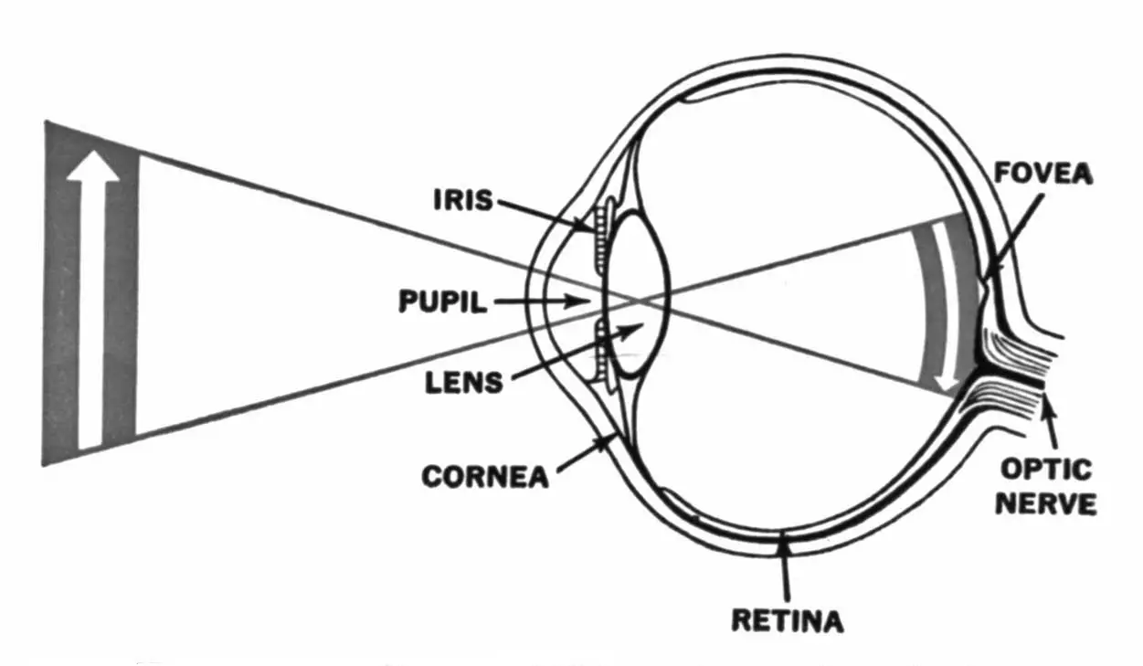
The article starts off by detailing the similarities between the human eye and a photographic lens. Both have an iris that opens and closes to regulate the amount of light that passes through it, both have a lens that focuses and inverts an image, and both have a light sensitive optical plane. In the case of a camera, a flat piece of film is what captures the image, but in our eye a curved surface made of light sensitive rods and cones send electrical signals to our brain, putting the signals together to form an image.
Like a camera lens, the eye’s iris widens as light is decreased and closes as light increases. But where a photographer would change to a faster film speed in low light the human eye has a similar ability of variable sensitivity through things called rods and cones. In the simplest terms, both rods and cones work together, but cones work singularly and are sensitive to bright lights and colors, whereas rods work in groups and are more sensitive to low light at the expense of color, so in a way, human vision starts off as a vibrant color film like Kodak Ektar 100 in daylight, and switches to a high sensitivity black and white film like TMax P3200 at night.
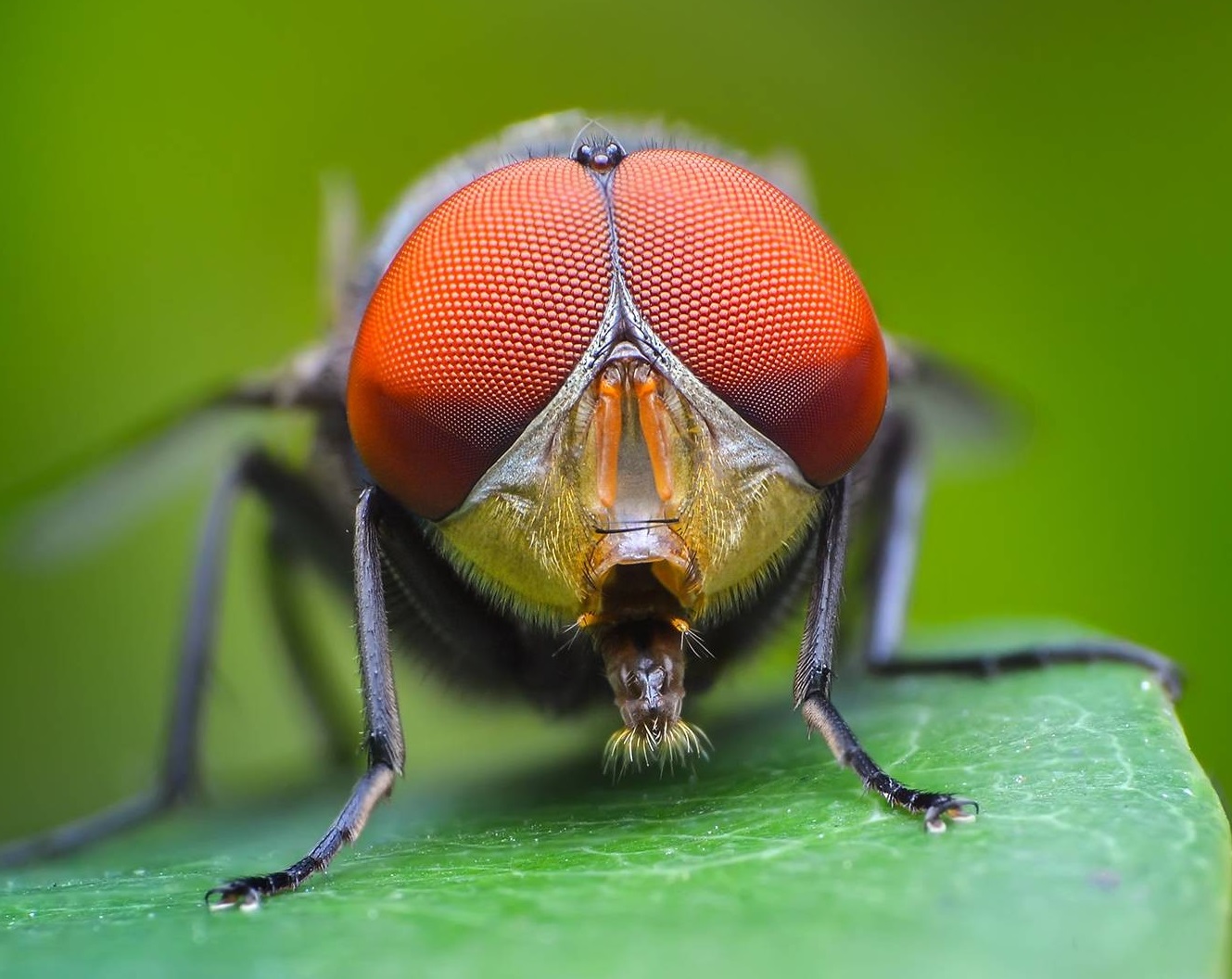
In the animal kingdom, not all eyes work like ours. The article briefly goes over how some species have eyes that are more similar to a pinhole camera that sacrifices detail for extreme depth, compound eyes like that of a housefly in which several images are pieces together to form one highly detailed image, and even that of the Copilia whose eye scans back and forth, sort of like a backwards Cathode Ray Tube in an old television.
Although our eyes and camera lenses share some similar principles, how they accomplish the act of converging light rays to form an image is very different. In a camera lens, focus is achieved by moving one or more lens groupings forward and aft. In the human eye, the entire eye changes from a more oval to circular shape. In a camera lens, spherical aberrations are corrected through multiple lens elements, in the human eye, the refracting elements are flatter in the middle and more rounder on the edges and also have variable light refracting properties in the center than the edges.
Chromatic aberrations, in which different wavelengths of light in the visible spectrum aren’t focused on the same point, are corrected in camera lenses via multiple elements and lens coatings, but in the human eye, the sensitivity of our rods and cones are dominant towards blue-green and yellow-red respectively. The article suggests an experiment to prove this in which if you look at two equally illuminated red and blue lights, the red light will seem brighter and harder to stare at in high intensity, but when the intensity decreases, the blue light will be easier to see in darkness than the red proving how our perception of color varies with intensity.
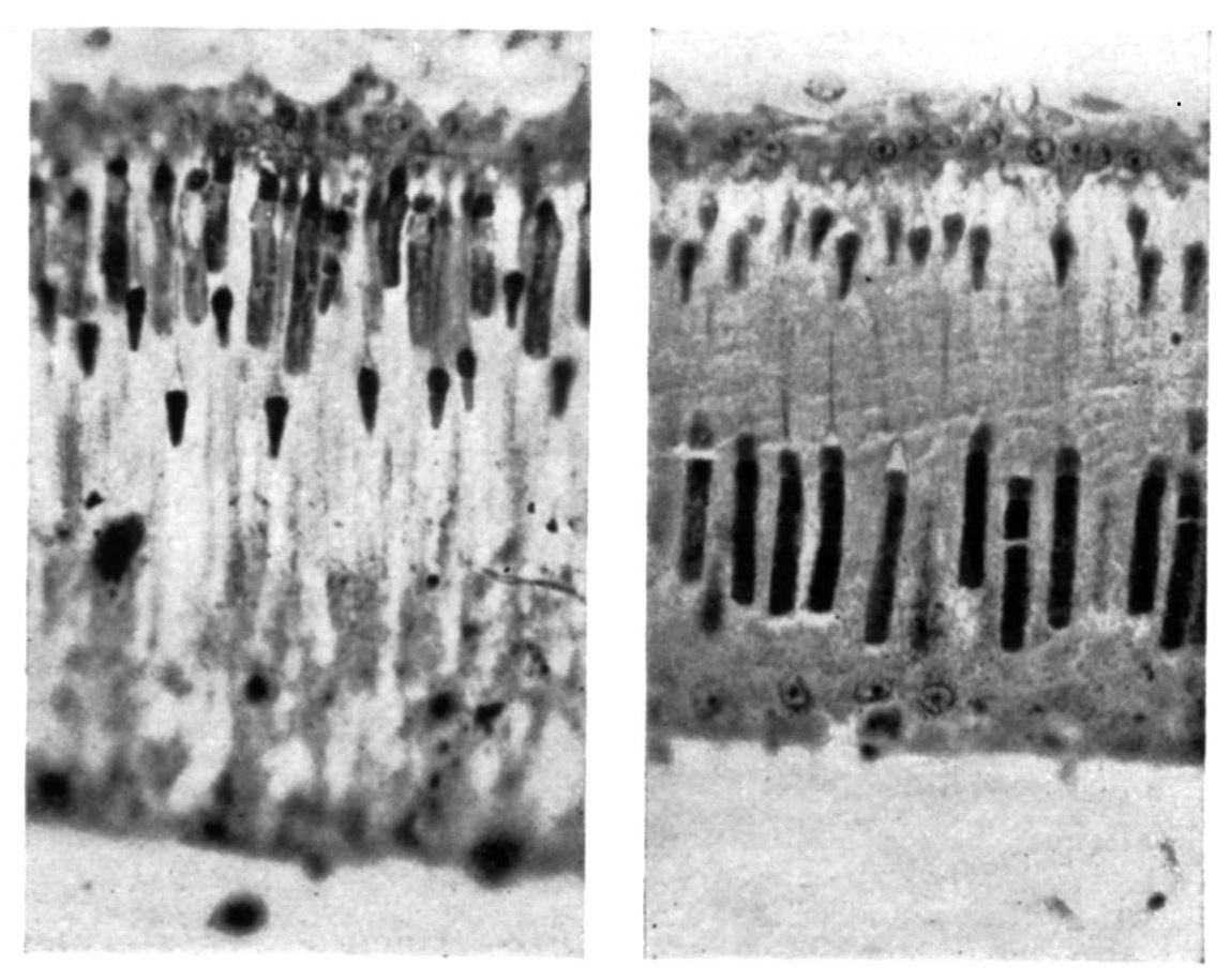
The article starts to go into some pretty off the wall discussions, at one point involving a German physiologist named Willy Kuehne and decapitated rabbits in which the eye was dissected after being exposed to bright vertical patterns to reveal a similar positive image on the back of the rabbit’s retina.
The discussion about the chemical makeup of the eye was less interesting to me, and sadly, the article never answers my question of what the f/stop or film speed of my eye is, but it is a fascinating read nonetheless.
I can’t help but wonder what kinds of conclusions have been reached in the 59 years since this article was published. How much of that research can be applied to photographic lenses today? Considering many manufacturers still use the same lens formulas that made up the Zeiss Tessar and Voigtländer Heliar a century ago are still being used today, suggest that not much has changed, but imagine if one day, a camera lens could be created that changed it’s shape to accommodate for focus, and also had lens elements made of material with different light refracting properties in different positions, or digital sensors that could vary ISO sensitivity within the same image.
All scans used with permission by Marc Bergman, 2020.
Wow. Hard to imagine being a photographer with just one eye. You are a dedicated man, that’s for sure.
That’s why I’ve never considered myself much of a photographer. I’m just a guy who really likes old cameras and the stories they tell! 🙂